Visualizing internal structures – penetrating and precise
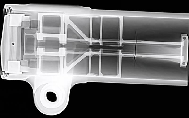
Radiographic testing is based on the varying permeability of materials to ionizing radiation. The component is irradiated with X-rays or gamma rays. The radiation is attenuated to varying degrees depending on material density, thickness, and internal irregularities.
Behind the component is a detector – such as film, a digital flat-panel detector, or an image plate system – that captures the transmitted residual radiation. Areas with higher material density absorb more radiation and appear brighter in the image, while areas with lower density or voids are displayed as darker.
If there is a defect inside the component, such as a shrinkage cavity, porosity, or bonding defect, this locally alters the radiation absorption. This creates a contrast difference in the captured image, allowing conclusions to be drawn about the location, size, and shape of the irregularity.
Image evaluation is performed using defined evaluation standards, reference specimens, or contractually agreed-upon criteria. Depending on the inspection task, different radiation sources, energy ranges, and exposure parameters are used to ensure sufficient image quality and inspection reliability.
X-ray inspection thus enables a two-dimensional representation of the internal condition of the component and is an established volumetric inspection method, particularly for castings, welds, and complex geometries.
